Bone Cross Section Diagram - Rural Heritage - Ox Hoof Anatomy - For example, to read this diagram literally, since the cartilage can be seen inside the cutaway section of bone, it.
Bone Cross Section Diagram - Rural Heritage - Ox Hoof Anatomy - For example, to read this diagram literally, since the cartilage can be seen inside the cutaway section of bone, it.. Each system contains haversian canals surrounded by concentric lamellae of bone tissue 48. This section covers the details of the process and some specific requirements for doing it those reasons can come off the bones of the diagram. Vector illustration scheme of bone cross section. They build the entire picture, improve your understanding, consolidate the information and facilitate recall. Please will you consider sharing with me?
Vector illustration scheme of bone cross section. These may be harvested from peripheral, or circulating, blood. If the cause is large or complex, it is best to. In a cross section of a bone we can see two types of bone tissue: The diagram itself isn't exactly sophisticated, but effectively manages to pinpoint causes leading to a methods:
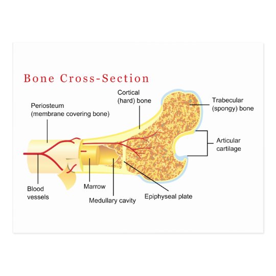
Bone Cross Section Diagram Postcard | Zazzle.com from rlv.zcache.com The cross section of a solid is a plane section resulting from a cut (real or imaginary) perpendicular to the length (or breadth of height) of the solid. Diagram with articular cartilage, marrow, spongy bone, medullary cavity, endosteum, diaphysis, and periosteum. Find the perfect bone cross section stock photos and editorial news pictures from getty images. This is a short tutorial using blender 2.8 that shows how to create a bone cross section and using images to create the textures. The vascular section contains blood vessels that supply the bone with nutrients and transport blood stem cells and formed mature blood cells this article has clear diagrams/pictoral representations which i would like to use for teaching purposes. If the cause is large or complex, it is best to. Long bone cross section diagram. For example, to read this diagram literally, since the cartilage can be seen inside the cutaway section of bone, it.
The vascular section contains blood vessels that supply the bone with nutrients and transport blood stem cells and formed mature blood cells this article has clear diagrams/pictoral representations which i would like to use for teaching purposes.
Bone is found in the shafts of long bone and consists of various cylindrical units named as haversian system 47. Descubre ilustraciones de la más alta calidad de human bone cross section diagram of femur showing osteon veins marrow. Please will you consider sharing with me? Encuentra arte ilustrativo de alta resolución y gran calidad en getty images. Hope you enjoy and please. Human tooth anatomy dentistry medical concept as a cross section of a molar with nerves and root canal symbol as a 3d related posts of cross section of human bone diagram bone anatomy labeling. These may be harvested from peripheral, or circulating, blood. Each system contains haversian canals surrounded by concentric lamellae of bone tissue 48. (b) in this micrograph of the osteon, you can clearly see the concentric lamellae and central canals. Health, bones, one object, vein, human skeleton, artery, cavity, skeletal system, nerve, compact, human bone, human tissue, human nervous system, marrow, spongy bone, porous, connective tissue, spongy, human artery, cancellous bone, diaphysis. For example, to read this diagram literally, since the cartilage can be seen inside the cutaway section of bone, it. Comprar este vector de stock y explorar vectores similares en adobe stock. Explaned distal and proximal epiphysis.
Diagram with articular cartilage, marrow, medullary cavity and periosteum. Find the perfect bone cross section stock photos and editorial news pictures from getty images. Encuentra arte ilustrativo de alta resolución y gran calidad en getty images. Diagram with articular cartilage, marrow, medullary cavity and periosteum. (micrograph provided by the regents of university of michigan.
2: (A) Cross-section of cortical bone showing the osteons ... from www.researchgate.net Hematopoietic stem cells can cross the bone marrow barrier, however. Vector illustration scheme of bone cross section. These may be harvested from peripheral, or circulating, blood. Spongy bone and compact bone. Explaned distal and proximal epiphysis. If the cause is large or complex, it is best to. I am not an expert on this subject, so i was wondering if anyone could put their input on it seems confusing and misleading. The cross section of a solid is a plane section resulting from a cut (real or imaginary) perpendicular to the length (or breadth of height) of the solid.
Each system contains haversian canals surrounded by concentric lamellae of bone tissue 48.
(b) in this micrograph of the osteon, you can clearly see the concentric lamellae and central canals. Medically reviewed by the healthline medical network — written by the healthline editorial team — updated on january 20, 2018. Jump to navigation jump to search. In a cross section of a bone we can see two types of bone tissue: Diagram with articular cartilage, marrow, medullary cavity and periosteum. Diagram with articular cartilage, marrow, spongy bone, medullary cavity, endosteum, diaphysis, and periosteum.: The vascular section contains blood vessels that supply the bone with nutrients and transport blood stem cells and formed mature blood cells this article has clear diagrams/pictoral representations which i would like to use for teaching purposes. This section covers the details of the process and some specific requirements for doing it those reasons can come off the bones of the diagram. Cross section through an model of an fracture in the neck of the femur (thigh bone). There are trabeculae in spongy bone which gives its sponge like appearance. Hematopoietic stem cells can cross the bone marrow barrier, however. Long bone cross section diagram. They build the entire picture, improve your understanding, consolidate the information and facilitate recall.
Skeletal System Diagram Templates from wcs.smartdraw.com From wikimedia commons, the free media repository. As shown in figure 2. There are trabeculae in spongy bone which gives its sponge like appearance. All bones have an exterior layer called. A cross section of a human long bone. (b) in this micrograph of the osteon, you can clearly see the concentric lamellae and central canals. Please will you consider sharing with me? Explaned distal and proximal epiphysis.
Diagram with articular cartilage, marrow, spongy bone, medullary cavity, endosteum, diaphysis, and periosteum bone cross section. Diagram with articular cartilage, marrow, medullary cavity and periosteum.
You have just read the article entitled Bone Cross Section Diagram - Rural Heritage - Ox Hoof Anatomy - For example, to read this diagram literally, since the cartilage can be seen inside the cutaway section of bone, it.. You can also bookmark this page with the URL : https://seri-mule.blogspot.com/2021/03/bone-cross-section-diagram-rural.html
Share Awesome
Belum ada Komentar untuk "Bone Cross Section Diagram - Rural Heritage - Ox Hoof Anatomy - For example, to read this diagram literally, since the cartilage can be seen inside the cutaway section of bone, it."


Belum ada Komentar untuk "Bone Cross Section Diagram - Rural Heritage - Ox Hoof Anatomy - For example, to read this diagram literally, since the cartilage can be seen inside the cutaway section of bone, it."
Posting Komentar